Rib Cage Muscles Diagram - - There is a printable worksheet available for download here so you can take the quiz with pen and paper.. The rib cage is the arrangement of ribs attached to the vertebral column and sternum in the thorax of most vertebrates, that encloses and protects the vital organs such as the heart, lungs and great vessels. This post is about rib cage. Further, there are two superior and two inferior processes meant for articulation with the neighbouring vertebra. It has clear front side and back planes. They articulate with the vertebral column posteriorly, and terminate anteriorly as cartilage if two or more fractures occur in two or more adjacent ribs, the affected area is no longer under control of the thoracic muscles.
It is formed by the vertebral column, ribs, and sternum and encloses the heart and lungs. Muscles that move the rib cage attach to the rib cage. Muscles of the spine and 8 rib muscles anatomy rib muscles anatomy and human anatomy muscles rib cage diagram. When the upper arm is lifted away from the torso, the the thick outer edge is the anterior wall of the axillary (armpit) region. All muscles that are attached to the human rib cage have the inherent potential to cause a breathing action.
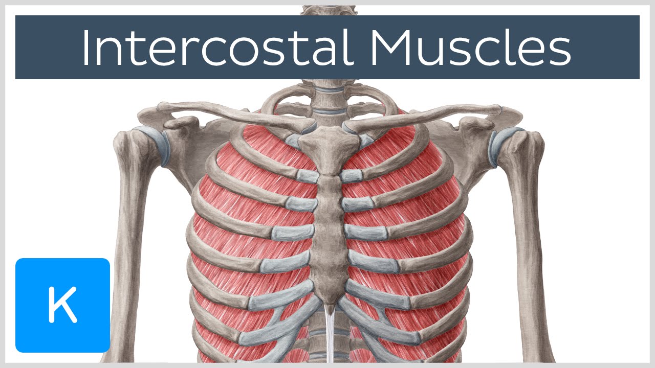
Intercostal Muscles - Function, Area & Course - Human ... from i.ytimg.com Muscles of the spine and 8 rib muscles anatomy rib muscles anatomy and human anatomy muscles rib cage diagram. The primary responsibilities of the ribcage involve protecting the thoracic visceral organs, enclosing the thoracic visceral organs, and is included in the general mechanics of the process of breathing. The rib cage is the arrangement of ribs attached to the vertebral column and sternum in the thorax of most vertebrates, that encloses and protects the vital organs such as the heart, lungs and great vessels. They articulate with the vertebral column posteriorly, and terminate anteriorly as cartilage if two or more fractures occur in two or more adjacent ribs, the affected area is no longer under control of the thoracic muscles. This is an online quiz called rib cage muscle diagram. The other attachment of these muscles is usually considered to be either superior or inferior to the rib attachment. The ribs joint as follows: It is one of six muscles that control the movements of the eye.
Muscles that move the rib cage attach to the rib cage.
Muscles of thorax, upper extremities, back and diaphragm are given connection by this cage. 2006 kia optima belt diagram. Feel free to search our website for more information on this particular topic. Each articulates with a thoracic vertebra. Rib cage diagram this summary post is displaying rib cage diagram. Anterior view of the lungs and ribcage in a transparent female torso stock illustration these pictures of this page are about:human anatomy rib cage muscles. Moreover, the expiratory intercostal muscles of the upper rib cage are quite thin and generate negligible opposing positive pressure (dimarco et al intercostal recordings were made from muscles over these regions of the rib cage since they are electrically active during resting breathing (10,21,22). When the upper arm is lifted away from the torso, the the thick outer edge is the anterior wall of the axillary (armpit) region. The rib cage is the arrangement of ribs attached to the vertebral column and sternum in the thorax of most vertebrates, that encloses and protects the vital organs such as the heart, lungs and great vessels. These rib muscles automatically get worked when you do bench presses, push ups and dips, but a few bonus exercises can help you really zero in for a more chiseled torso. All muscles that are attached to the human rib cage have the inherent potential to cause a breathing action. Posted on december 22, 2018december 22, 2018. It encloses and protects the heart and lungs.
The function of the rib cage is to filter the blood it receives, processing the blood. Posted on december 22, 2018december 22, 2018. Muscle spasms located in the rib cage are often observed in people who strain or overwork their upper body muscles. So what parts of the rib cage show up on the surface? Best viewed on 1280 x 768 px resolution in any modern browser.
The Breath of Life (part 2) — Alexander Technique in ... from static1.squarespace.com It is formed by the vertebral column, ribs, and sternum and encloses the heart and lungs. The accompanying diagram reveals the actions of the muscles in this pose. The primary responsibilities of the ribcage involve protecting the thoracic visceral organs, enclosing the thoracic visceral organs, and is included in the general mechanics of the process of breathing. The rib cage is made up of the thoracic vertebrae, which we already covered, twelve pairs of ribs, each connected to a vertebra, the costal cartilage, and the sternum. The ribs are a set of twelve paired bones which form the protective 'cage' of the thorax. It is one of six muscles that control the movements of the eye. These muscles may be located anteriorly, posteriorly, and/or laterally. The function of the rib cage is to filter the blood it receives, processing the blood.
The two muscles which comprise the intermediate muscle group are the serratus posterior inferior, and the serratus posterior superior.
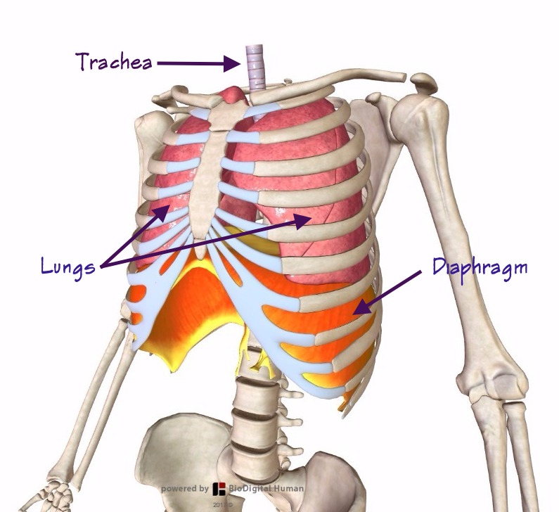
The function of the rib cage is to filter the blood it receives, processing the blood. Each articulates with a thoracic vertebra. Muscles that helpful in expanding the thoracic cavity are called the inspiratory muscles because they help in inhalation, while those that compress the thoracic cavity are called expiratory. Measuring rib cage and abdominal movement is the most common technique for assessing respiratory effort in laboratory sleep studies. It provides a strong framework onto which the muscles of the shoulder girdle, chest, upper abdomen and back can attach. This is an online quiz called rib cage muscle diagram. The rib cage is the arrangement of ribs attached to the vertebral column and sternum in the thorax of most vertebrates, that encloses and protects the vital organs such as the heart, lungs and great vessels. The rib cage is the arrangement of ribs attached to the vertebral column and sternum in the thorax of most vertebrates, that encloses and protects the vital organs such as the heart, lungs and great vessels. During normal breathing, the major inspiratory muscles produce rib cage expansion and a downward movement of the diaphragm. As you inhale, the muscles in between the ribs lift the rib cage up, allowing the lungs to expand. Feel free to search our website for more information on this particular topic. The primary responsibilities of the ribcage involve protecting the thoracic visceral organs, enclosing the thoracic visceral organs, and is included in the general mechanics of the process of breathing. So what parts of the rib cage show up on the surface?
This is an online quiz called rib cage muscle diagram. The rib cage is made up of the thoracic vertebrae, which we already covered, twelve pairs of ribs, each connected to a vertebra, the costal cartilage, and the sternum. It encloses and protects the heart and lungs. Learn vocabulary, terms and more with flashcards, games and other study tools. This post is about rib cage.
Rib Cage Diagram With Organs - Human Anatomy Body from www.anatomylibrary99.com Start studying rib cage muscles. It is one of six muscles that control the movements of the eye. The rib cage is composed by sternum, costal cartilages, and ribs connected to the thoracic vertebrae. These rib muscles automatically get worked when you do bench presses, push ups and dips, but a few bonus exercises can help you really zero in for a more chiseled torso. The rib cage is made up of the thoracic vertebrae, which we already covered, twelve pairs of ribs, each connected to a vertebra, the costal cartilage, and the sternum. The ribs joint as follows: The other attachment of these muscles is usually considered to be either superior or inferior to the rib attachment. Diversitech condensate pump wiring diagram.
Muscles that move the rib cage attach to the rib cage.
Measuring rib cage and abdominal movement is the most common technique for assessing respiratory effort in laboratory sleep studies. The two muscles which comprise the intermediate muscle group are the serratus posterior inferior, and the serratus posterior superior. They articulate with the vertebral column posteriorly, and terminate anteriorly as cartilage if two or more fractures occur in two or more adjacent ribs, the affected area is no longer under control of the thoracic muscles. When you exhale, the rib cage moves down again, squeezing the air. So what parts of the rib cage show up on the surface? The rib cage is composed by sternum, costal cartilages, and ribs connected to the thoracic vertebrae. On the dorsal side there is a neural spine. Start studying rib cage muscles. These rib muscles automatically get worked when you do bench presses, push ups and dips, but a few bonus exercises can help you really zero in for a more chiseled torso. Muscles that helpful in expanding the thoracic cavity are called the inspiratory muscles because they help in inhalation, while those that compress the thoracic cavity are called expiratory. The following general rules regarding actions can be. In humans, the rib cage, also known as the thoracic cage. Muscle spasms located in the rib cage are often observed in people who strain or overwork their upper body muscles.
This is an online quiz called rib cage muscle diagram rib cage muscles. Muscles that helpful in expanding the thoracic cavity are called the inspiratory muscles because they help in inhalation, while those that compress the thoracic cavity are called expiratory.


0 Comments